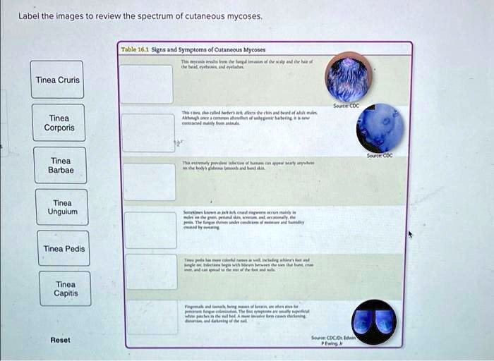
Label the images to review the spectrum of cutaneous mycoses – Labeling images to review the spectrum of cutaneous mycoses stands as a cornerstone in the field of dermatology, providing a critical foundation for accurate diagnosis and effective management of these common infections. This comprehensive guide delves into the significance of consistent and precise image labeling, exploring its benefits, criteria, and implications for the diagnosis and treatment of cutaneous mycoses.
Through a detailed examination of the spectrum of cutaneous mycoses, including superficial mycoses, cutaneous candidiasis, dermatophytosis, Malassezia furfur infections, and pityriasis versicolor, this guide equips readers with a thorough understanding of the clinical manifestations, epidemiology, and treatment options associated with each type.
1. Introduction to Cutaneous Mycoses

Cutaneous mycoses, also known as skin mycoses, are infections of the skin, hair, and nails caused by fungi. They are a common health problem worldwide, affecting individuals of all ages and backgrounds. Accurate labeling of images is crucial for reviewing the spectrum of cutaneous mycoses, as it enables consistent and effective analysis of clinical manifestations and facilitates accurate diagnosis and management.
2. Image Labeling for Cutaneous Mycoses

Images play a vital role in the review of cutaneous mycoses, providing a visual representation of the clinical manifestations and aiding in the diagnosis and management of these infections. Consistent and accurate image labeling is essential for effective analysis, as it allows for the systematic organization and retrieval of images based on specific criteria.
Criteria for Image Labeling
- Type of cutaneous mycosis
- Clinical presentation (e.g., morphology, distribution, color)
- Anatomical location
- Treatment response
- Patient demographics (e.g., age, sex, medical history)
3. Spectrum of Cutaneous Mycoses: Label The Images To Review The Spectrum Of Cutaneous Mycoses

Superficial Mycoses, Label the images to review the spectrum of cutaneous mycoses
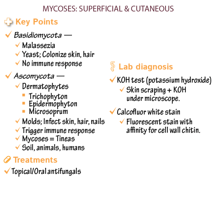
Superficial mycoses are caused by fungi that invade only the outermost layer of the skin, known as the stratum corneum. They typically present with scaly, erythematous patches on the skin.
Cutaneous Candidiasis
Cutaneous candidiasis is caused by Candida species, which are commonly found on the skin and mucous membranes. It can manifest as a variety of clinical presentations, including intertrigo, diaper rash, and paronychia.
Dermatophytosis
Dermatophytosis, commonly known as ringworm, is caused by dermatophyte fungi. It affects the skin, hair, and nails, leading to a characteristic annular rash with raised borders and central clearing.
Malassezia Furfur Infections
Malassezia furfur is a lipophilic yeast that can cause a range of skin conditions, including pityriasis versicolor and seborrheic dermatitis. Pityriasis versicolor presents as hypopigmented or hyperpigmented macules on the trunk and proximal extremities.
4. Case Studies and Image Gallery
Case studies and images are invaluable for illustrating the clinical manifestations and diagnostic challenges associated with cutaneous mycoses. A series of case studies with high-quality images and detailed annotations will be presented, showcasing the spectrum of cutaneous mycoses and providing insights into their diagnosis and management.
FAQ Corner
What are the benefits of using images for reviewing the spectrum of cutaneous mycoses?
Images provide a visual representation of the clinical manifestations of cutaneous mycoses, allowing for more accurate diagnosis and assessment of disease severity.
Why is consistent and accurate image labeling important for effective analysis?
Consistent and accurate image labeling ensures that images can be easily searched, retrieved, and compared, facilitating the identification of patterns and trends in the presentation of cutaneous mycoses.
What are some of the criteria that should be considered when labeling images of cutaneous mycoses?
Criteria for labeling images of cutaneous mycoses include the type of mycosis, anatomical location, clinical presentation, and treatment response.